Permeability of a biological membrane
Animal cells have a fluid membrane. This allows them some degree of fluidity, but it can also extract a price - they cannot withstand an increase in cell volume.
Plant cells can - they have rigid cellulose walls and can develop a turgor pressure to counter any hydrostatic pressure increase that causes fluid to flow into the cells.
Animal cells can't, so if fluid floods into the animal cell, it will rapidly swell and burst. This practical is all about that swelling - what is called "lysis". We are going to study that process, using red blood cells (RBCs), for two main reasons:
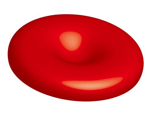
First, unlike almost all other cells in the animal's body, RBCs have a small capacity to swell. They have a doughnut shape as you can see in this image from Wikimedia:

This means they can increase their volume to become more round, and only then reach the limit of their swelling capacity and burst. As this takes a little time (and that is what you will measure in this practical class), you can therefore measure the time taken for the RBCs to burst (to lyse - or haemolysis, since the lysis involves blood cells).
Second, they (and white blood cells) are the ones most directly facing fluid challenges as fluid moves into and out of the cardiovascular system. These are also the cells first challenged if you lose significant blood volume (a large wound) and need a transfusion. So we need to know that the fluid we put into the body in that case won't cause cells to lyse and die.
Note that while we have talked about cells swelling and bursting (lysis), shrinking of cells (called creation) as excess fluid moves out of them is also a bad thing. It means cellular organelles are no longer in ideal positions to interact with each other - e.g., for the proteins produced on ribosomes to be appropriately packaged and delivered to the Golgi apparatus for modifications to make them the finished product for use, etc.
You can see the lysis and creation of RBCs in this YouTube video below:
We will use a very simple principle here:
- If cells are intact in a solution, then they will float around and impede the path of light you try to shine through the solution.
- Whereas, if cells are lysed in a solution, then the cell debris will be scattered and offer less of an impediment for light to be seen through the solution.
That's it. Measure how long it takes for RBCs placed in a solution to lyse by measuring how long it takes for you to be able to see a light bulb on the other side of the test tube.
Below is a simulation of what the two end points would look like - when the RBCs are intact and light can't get through easily, and when the RBCs are lysed, and you can see the light bulb through the solution.
In the window below, each of the two test tubes has previously had some RBCs put into a solution in the tube. You can move the test tubes around with your mouse to be in front of the light bulb on the right of the simulation and compare how they look.
First, move Tube 1 to the light on the right.
- This is how it looks when the RBCs are intact and floating around to block the path of light.
Now move Tube 2 to the light on the right.
- Notice that the bulb is clearly visible. This means the RBCs have undergone lysis (i.e., they have swollen and burst). The cell fragments have all settled to the bottom, while the cell organelles are floating around and not impeding the path of light.
- We can therefore see the filament of the light bulb much more clearly.
In this class, you will be manipulating the type and concentration of the particles of the solution to see how this influences the time taken for lysis to occur.
