How we perceive colours
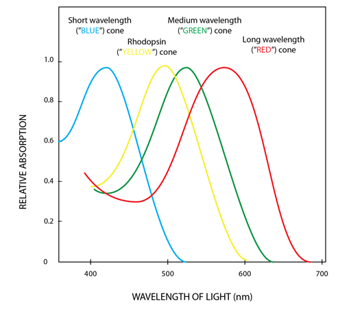
We have photoreceptors called cones which have photopigment molecules that differ from those in the rod photoreceptors (that we depend on for night vision), and that also differ from each other in one of three classes. The three cone photopigments (only one found in any one cone) contain an opsin apoprotein, covalently linked to 11-cis-hydroretinal, which is the light absorbing molecule. Slight differences in the apoprotein mean that there are differences between the cones as to their spectral sensitivities (i.e., the range of wavelengths of light at which they best absorb energy). The cones are termed according to the range of wavelengths of the peaks of their spectral sensitivities: short (S), medium (M), and long (L) cones.
These differences allow us to discriminate between different wavelengths of light. However, it's important to note that these three types do not correspond well to particular specific colours. Rather, as Wikipedia notes, the perception of colour is achieved by a complex process that starts with the differential output of these cells in the retina, and it will be finalized in the visual cortex and associative areas of the brain. Thus, while you may commonly hear the cone receptors being termed red receptors, or green receptors or blue receptors, colour is a percept of the brain, not of the retina.

Testing colour blindness
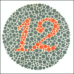
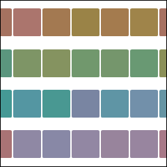
In this section you can conduct two types of colour vision (more strictly, colour deficit) measurements: first, the common Ishihara plates that are more biased to detecting red/green colour blindness, and then a second simulation, the Farnsworth-Munsell Hue Colour Blindness test. The latter is much more extensive in allowing you comparison of your colour perception data to a normative database to evaluate your colour vision.
Further testing
For more tests relating to visual acuity, contrast acuity and colour vision, head over to:
https://www.zeiss.com/vision-care/en/eye-health-and-care/zeiss-online-vision-screening-check.html


